Marburg Virus Diagram : Clinical Aspects Of Marburg Hemorrhagic Fever Future Virology / Gastrointestinal distress, including watery diarrhea, nausea, and cramping, often around three days after symptoms appear.
Marburg Virus Diagram : Clinical Aspects Of Marburg Hemorrhagic Fever Future Virology / Gastrointestinal distress, including watery diarrhea, nausea, and cramping, often around three days after symptoms appear.. In the early course of the disease, clinical diagnosis of mvd is difficult to distinguish from other tropical febrile illnesses, because of the similarities in the clinical symptoms. Like ebola, marburg virus disease can cause severe hemorrhaging that leads to shock, organ failure, or death. Molecular structures and visual images of the proteins of marburg virus are essential for the development of antiviral drugs. Comments and questions to info@ncbi.nlm.nih.gov Marburg virus disease (mvd), formerly known as marburg haemorrhagic fever, is a severe, often fatal illness in humans.
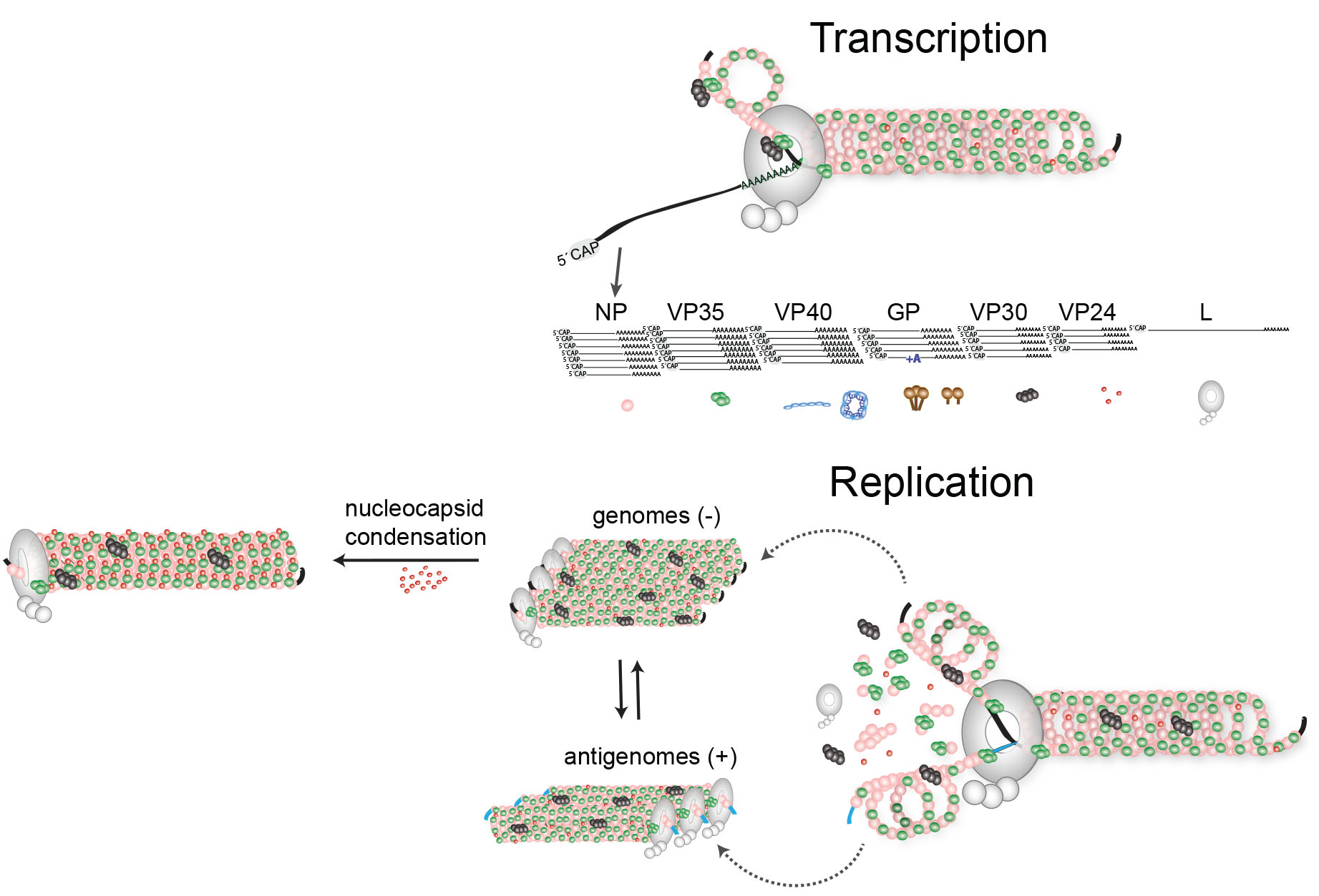
This diagram shows how ebola virus. As you can see in figure 2, the green helical structure is the genomic rna surrounded by polymer of nucleoproteins (np). Analyzing such images sheds light on the structure of the virus and the mechanisms by which it is assembled. (d) topology diagram of marburg virus vp24, with secondary structure elements sequentially numbered and colored from n to c as described for panels a to c. In addition, exposure to an infected human is high risk factor.
Diagnostics For Filovirus Detection Impact Of Recent Outbreaks On The Diagnostic Landscape Abstract Europe Pmc from europepmc.org Analyzing such images sheds light on the structure of the virus and the mechanisms by which it is assembled. As you can see in figure 2, the green helical structure is the genomic rna surrounded by polymer of nucleoproteins (np). Crystal structure of the marburg virus nucleoprotein core domain chaperoned by a vp35 peptide reveals a conserved drug target for filovirus j virol. Bacteria multiply by the billions. (d) topology diagram of marburg virus vp24, with secondary structure elements sequentially numbered and colored from n to c as described for panels a to c. Marburg virus is a hemorrhagic fever virus of the filoviridae family of viruses and a member of the species marburg marburgvirus, genus marburgvirus.marburg virus (marv) causes marburg virus disease in humans and other primates, a form of viral hemorrhagic fever. Comments and questions to info@ncbi.nlm.nih.gov This protein mediates infection by binding to the viral envelope glycoprotein.
The marburg virus contains seven structural proteins.
The world health organization (who) rates it as a risk group 4 pathogen. Mvd is a viral hemorrhagic fever (vhf), and the clinical symptoms are indistinguishable from ebola virus disease (evd). Risk factors include exposure to african green monkeys and certain bats; In the early course of the disease, clinical diagnosis of mvd is difficult to distinguish from other tropical febrile illnesses, because of. Bacteria multiply by the billions. This diagram shows how ebola virus. Mvd case fatality rates have been as high as 80 to 90 percent. Marburg virus was first introduced under this name in 1967.6 in 2005, the virus name was changed to lake victoria marburgvirus, which unfortunately was the same spelling as the species. Marburg virus is a hemorrhagic fever virus of the filoviridae family of viruses and a member of the species marburg marburgvirus, genus marburgvirus.marburg virus (marv) causes marburg virus disease in humans and other primates, a form of viral hemorrhagic fever. Marburg virus disease is caused by viruses that produce symptoms of fever, chills, headaches and muscle aches early in the disease; Marburg virus structure and transmission. It is unknown how marburg virus first transmits from its animal host to humans; The marburg virus does not contain the polyadenylation sequence that is found in the ebola gp gene.
One key protein in the marburg virus life cycle is … In addition, exposure to an infected human is high risk factor. The marburg virus contains seven structural proteins. Risk factors include exposure to african green monkeys and certain bats; Gastrointestinal distress, including watery diarrhea, nausea, and cramping, often around three days after symptoms appear.
Research Muhlberger Lab from www.bu.edu Scientists have determined the structure of a critical protein from the marburg virus, a close cousin of ebola virus. Marburg, which is in the same family as the virus that causes ebola, was detected less than two months after guinea declared an end to an ebola outbreak that erupted earlier this year. As you can see in figure 2, the green helical structure is the genomic rna surrounded by polymer of nucleoproteins (np). Marburg virus disease (mvd), formerly known as marburg haemorrhagic fever, is a severe, often fatal illness in humans. This protein mediates infection by binding to the viral envelope glycoprotein. The marburg virus does not contain the polyadenylation sequence that is found in the ebola gp gene. Comments and questions to info@ncbi.nlm.nih.gov In humans, marburgviruses are responsible for marburg virus disease (mvd), a zoonotic disease that is characterized by high fever, malaise, nausea, vomiting, diarrhea, skin rash, and hemorrhage (bleeding).
Comments and questions to info@ncbi.nlm.nih.gov
Risk factors include exposure to african green monkeys and certain bats; The world health organization (who) rates it as a risk group 4 pathogen. The soviet union experimented with mvd toward the end of the cold war, in the 1980s and 1990s, in an attempt to develop a potent biological weapon. Marburg virus is a deadly pathogen that causes marburg disease a severe viral hemorrhagic fever, named after the city in germany, where the first outbreak. As you can see in figure 2, the green helical structure is the genomic rna surrounded by polymer of nucleoproteins (np). For the marburg virus to infect the host's cell an essential element is needed. Analyzing such images sheds light on the structure of the virus and the mechanisms by which it is assembled. This diagram shows how ebola virus. The virus is considered to be extremely dangerous. It is unknown how marburg virus first transmits from its animal host to humans; As with most of griyo's viruses, marburg is named for a human/animal virus. Process flow diagram for mrna drug substance production (aka. The marburg virus does not contain the polyadenylation sequence that is found in the ebola gp gene.
In the early course of the disease, clinical diagnosis of mvd is difficult to distinguish from other tropical febrile illnesses, because of the similarities in the clinical symptoms. The potential for the marburg virus to spread far and wide means we need to stop it in its tracks marburg case fatality rates have varied from 24% to 88% in past outbreaks, depending on virus. Marburg virus structure and transmission. The marburgvirus genus includes two viruses. As you can see in figure 2, the green helical structure is the genomic rna surrounded by polymer of nucleoproteins (np).
Rcsb Pdb 5uqy Crystal Structure Of Marburg Virus Gp In Complex With The Human Survivor Antibody Mr78 from files.rcsb.org In humans, marburgviruses are responsible for marburg virus disease (mvd), a zoonotic disease that is characterized by high fever, malaise, nausea, vomiting, diarrhea, skin rash, and hemorrhage (bleeding). Along with ebola virus, marburg virus causes a severe and highly fatal haemorrhagic. Analyzing such images sheds light on the structure of the virus and the mechanisms by which it is assembled. Marburg virus diagram / research muhlberger lab. Marburg virus causes symptoms that come on suddenly and become increasingly severe. Tried, tested, trusted and affordable for all qpcr needs. Process flow diagram for mrna drug substance production (aka. Marburg virus is a deadly pathogen that causes marburg disease a severe viral hemorrhagic fever, named after the city in germany, where the first outbreak.
These viruses cause similar diseases and are some of the deadliest pathogens.
Risk factors include exposure to african green monkeys and certain bats; Marv was first isolated in 1967, following an outbreak of hemorrhagic illness. Marburg virus causes symptoms that come on suddenly and become increasingly severe. Mvd is a viral hemorrhagic fever (vhf), and the clinical symptoms are indistinguishable from ebola virus disease (evd). The viral fragment is pleomorphic, and may appear in the shape of a 6, a u, or a circle, and it is contained within a lipid. Marburg virus disease (mvd), formerly known as marburg haemorrhagic fever, is a severe, often fatal illness in humans. Molecular structures and visual images of the proteins of marburg virus are essential for the development of antiviral drugs. Marburg virus is a deadly pathogen that causes marburg disease a severe viral hemorrhagic fever, named after the city in germany, where the first outbreak. One key protein in the marburg virus life cycle is … Marburg virus is the causative agent of marburg virus disease (mvd), a disease with a case fatality ratio of up to 88%, but can be much lower with good patient care. Crystal structure of the marburg virus nucleoprotein core domain chaperoned by a vp35 peptide reveals a conserved drug target for filovirus j virol. Marburg virus was first recognized in 1967, when outbreaks of hemorrhagic fever occurred simultaneously in laboratories in marburg and frankfurt, germany and in belgrade, yugoslavia (now serbia). Gastrointestinal distress, including watery diarrhea, nausea, and cramping, often around three days after symptoms appear.


0 Komentar